Fluoreszcens nanorészecske követéssel kivitelezett mérések standardizálása
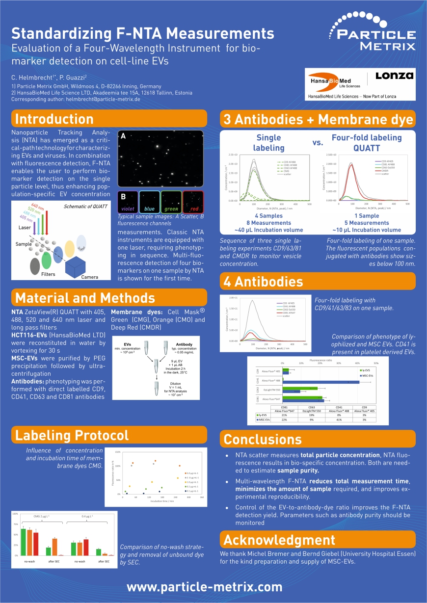
A fluoreszcens detektálással kombinált nanorészecske követés (F-NTA: Fluorescent detection-Nanoparticle Tracking Analysis) lehetővé teszi az extracelluláris vezikulák biomarker alapú vizsgálatát. A bemutatott munkában a ZetaView QUATT készülék használatával először végeztek multifluoreszcens detektálást négy különböző biomarker alkalmazá-sával ugyanazon a mintán. Ez a gyakorlatban csökkenti a méréshez szükséges időt és a minta mennyiségét, illetve növeli a kísérlet reprodukálhatóságát.
Standardizing F-NTA Measurements
Evaluation of a Four-Wavelenght Instrument for biomarker detection on cell-line EVs
C.Helmbrecht1, P. Guazzi2
1) Particle Metrix GmbH, Wildmoos 4, D-82266 Inning, Germany
2) HansaBioMed Life Science LTD, Akadeemia tee 15A, 12618 Tallinn, Estonia Corresponding author: helmbrecht@particle-metrix.de
Introduction
Nanoparticle Tracking Analysis (NTA) has emerged as a critical-path technology for characterizing EVs and viruses. In combination with fluorescence detection, F-NTA enables the user to perform biomarker detection on the single particle level, thus enhancing population-specific EV concentration measurements. Classic NTA instruments are equipped with one laser, requiring phenotyping in sequence. Multi-fluorescence detection of four biomarkers on one sample by NTA is shown for first time.
Materials and Methods
NTA ZetaView(R) QUATT with 405, 488, 520 and 640 nm laser and long pass filters
HCT116-EVs (HansaBioMed LTD) were reconstituted in water by vortexing for 30s
MSC-EVs were purified by PEG precipitation followed by ultracentrifugation.
Antibodies: phenotyping was performed with direct labelled CD9, CD41, CD63, and CD81 antibodies
Membrane dyes: Cell Mask® Green (CMG), Orange (CMO) and Deep Red (CMDR)
Labeling Protocol
Influence of concentration and incubation time of membrane dyes CMG. (poster figure) Comparison of no-wash strategy and removal of unbound dye by SEC. (poster figure)
3 Antibodies + Membrane dye
Sequence of three singre labeling experiments CD9/63/81 and CMDR to monitor vesicle concentration. (poster figure)
Four-fold labeling of one sample. The fluorescent populations conjugated with antibodies show siezs below 100 nm. (poster figure)
4 Antibodies
Four-fold labeling with CD9/41/63/83 on one sample. (poster figure)
Comparison of phenotype of lyophilized and MSC EVs. CD41 is present in plateled derived EVs. (poster figure)
Conclusions:
- NTA scatter measures total particle concentration. NTA fluorescence results in bio-specific concentration. Both are needed to estimate sample purity.
- Multi-wavelenght F-NTA reduces total measurement time, minimizes the amount of sample required, and improves experimental reproducibility.
- Control of the EV-to-antibody-dye ratio improves the F-NTA detection yield. Parameters such as antibody purity should be monitored.
Acknowledgement
We thank Michel Bremer and Bernd Giebel (University Hospital Essen) for the kind preparation and supply of MSC-EVs.

hozzászólások (0)
Még nincs hozzászólás. Legyen Ön az első!